Πέμπτη 4 Φεβρουαρίου 2021

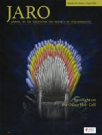
Residual Hair Cell Responses in Electric-Acoustic Stimulation Cochlear Implant Users with Complete Loss of Acoustic Hearing After Implantation

xlomafota13 shared this article with you from Inoreader

10162.jpg

Abstract

Changes in cochlear implant (CI) design and surgical techniques have enabled the preservation of residual acoustic hearing in the implanted ear. While most Nucleus Hybrid L24 CI users retain significant acoustic hearing years after surgery, 6–17 % experience a complete loss of acoustic hearing (Roland et al. Laryngoscope. 126(1):175-81. (2016), Laryngoscope. 128(8):1939-1945 (2018); Scheperle et al. Hear Res. 350:45-57 (2017)). Electrocochleography (ECoG) enables non-invasive monitoring of peripheral auditory function and may provide insight into the pathophysiology of hearing loss. The ECoG response is evoked using an acoustic stimulus and includes contributions from the hair cells (cochlear microphonic—CM) as well as the auditory nerve (auditory nerve neurophonic—ANN). Seven Hybrid L24 CI users with complete loss of residual hearing months after surgery underwent ECoG measures before and after loss of hearing. While significant reductions in CMs were evident after hearing loss, all participants had measurable CMs despite having no measurable acoustic hearing. None retained measurable ANNs. Given histological data suggesting stable hair cell and neural counts after hearing loss (e.g., Quesnel et al. Hear Res. 333:225-234. (2016)), the loss of ECoG and audiometric hearing may reflect reduced synaptic input. This is consistent with the theory that residual CM responses coupled with little to no ANN responses reflect a "disconnect" between hair cells and auditory nerve fibers (Fontenot et al. Ear Hear. 40(3):577-591. 2019). This "disconnection" may prevent proper encoding of auditory stimulation at higher auditory pathways, leading to a lack of audiometric responses, even in the presence of viable cochlear hair cells.

View on the web

Salivary Duct Carcinoma With Rhabdoid Features—No or Aberrant Expression of E-cadherin and Genetic Changes in CDH1: Immunohistochemical and Genetic Analyses of 17 Cases

xloma.fota13 shared this article with you from Inoreader

SocialThumb.00000478.DC.jpeg

Salivary duct carcinoma is a relatively uncommon malignancy of the salivary glands; however, it frequently occurs as a carcinomatous component of carcinoma ex pleomorphic adenoma. We previously reported salivary duct carcinoma with rhabdoid features (SDCRF) as an extremely rare subtype of salivary duct carcinoma, and that it occurred as a salivary counterpart of pleomorphic lobular carcinoma of the breast (PLCB). We collected new cases of SDCRF for this study, in which we examined a total of 17 cases immunohistochemically and genetically. As it is known that PLCB exhibits loss of or aberrant E-cadherin expression and carries nonsense/missense mutations in or deletion of the CDH1 gene, we examined the CDH1 gene status of our SDCRF cases. All of the examined SDCRF cases involved the diffuse proliferation of large ovoid cells with eosinophilic cytoplasm and eccentric nuclei, which displayed reduced cell-cell adhesion. Most cases were positive for pan-cytokeratin, androgen receptor, gross cystic disease fluid protein-15, SWI/SNF-related matrix-associated actin-dependent regulator of chromatin subfamily B member 1, and WI/SNF-related matrix-associated actin-dependent regulator of chromatin subfamily A member 4, whereas they were negative for vimentin. No and decreased/cytoplasmic E-cadherin expression was ob served in 11 and 4 of 17 cases, respectively, whereas no and decreased/cytoplasmic β-catenin expression were observed in 10 and 5 of 17 cases, respectively. Among the 11 cases that could be genetically analyzed, a nonsense mutation (1 case), missense mutations (6 cases), and insertions (1 case) were detected in the CDH1 gene. In conclusion, we propose that SDCRF is the salivary counterpart of PLCB due to its morphology and immunophenotype, and the genetic status of CDH1. This study was approved by the Institutional Review Board of Shizuoka General Center (SGHIRB#2019007) and Hamamatsu University School of Medicine (20-011). All subjects signed informed consent forms. Written informed consent for the publication of clinical details and/or clinical images was obtained from the patients. A copy of the consent form is available for review by the Editor of this journal. K.K.: designed and drafted the manuscript. K.K. and M.S.: made the histopathologic diagnoses. M.M., K.Y., S.B., T.O., and H.I.: found the cases and collected the clinical data. H. Yamamoto, T.I., and C.T.: performed the immunohistochemistry. H. Yamada and K.I.: performed the genetic analysis. H.S. and M.S.: supervised this manuscript. Conflicts of Interest and Source of Funding: Supported in part by a Grant-in-Aid for the Medical Research Support Project of Shizuoka Prefectural Hospital Organization in 2019 (to K.K.), as well as by AMED (20ck0106554) (to H.S.) and SRF (to H.S.). The authors have disclosed that they have no significant relationships with, or financial interest in, any commercial companies pertaining to this article. Correspondence: Kimihide Kusafuka, DDS, PhD, Department of Pathology, Shizuoka General Hospital, 4-27-1 Kita-Ando, Aoi-ku, Shizuoka City 420-8527, Shizuoka Prefecture, Japan (e-mail: k-kusafuka@i.shizuoka-pho.jp). Copyright © 2021 Wolters Kluwer Health, Inc. All rights reserved.
View on the web

A Distinctive Adnexal (Usually Paratubal) Neoplasm Often Associated With Peutz-Jeghers Syndrome and Characterized by STK11 Alterations (STK11 Adnexal Tumor): A Report of 22 Cases

xloma.fota13 shared this article with you from Inoreader

SocialThumb.00000478.DC.jpeg

We describe 22 examples of a novel, usually paratubal, adnexal tumor associated with Peutz-Jeghers syndrome in nearly 50% of cases that harbored STK11 alterations in all tested (n=21). The patients ranged from 17 to 66 years (median=39 y) and the tumors from 4.5 to 25.5 cm (median=11 cm). Most (n=18) were paratubal, wi th metastases noted in 11/22 (50%) and recurrences in 12/15 (80%). Morphologically, they were characterized by interanastomosing cords and trabeculae of predominantly epithelioid cells, set in a variably prominent myxoid to focally edematous stroma, that often merged to form tubular, cystic, cribriform, and microacinar formations, reminiscent of salivary gland-type tumors. The tumor cells were uniformly atypical, often with prominent nucleoli and a variable mitotic index (median=9/10 HPFs). The tumors were usually positive to a variable extent for epithelial (CAM5.2, AE1/AE3, cytokeratin 7), sex cord (calretinin, inhibin, WT1), and mesothelial (calretinin, D2-40) markers, as well as hormone receptors. PAX8, SF1, and GATA-3 were rarely positive, while claudin-4, FOXL2, and TTF-1 were consistently negative. All sequenced tumors (n=21) harbored alterations in STK11, often with a loss of heterozygosity event. There were no other recurrently mutated genes. Recurrent copy number alterat ions included loss of 1p and 11q, and gain of 1q, 15q, and 15p. Despite an extensive morphologic, immunohistochemical, and molecular evaluation, we are unable to determine with certainty the histogenesis of this unique tumor. Wolffian, sex cord stromal, epithelial, and mesothelial origins were considered. We propose the term STK11 adnexal tumor to describe this novel entity and emphasize the importance of genetic counseling in these patients as a significant number of neoplasms occur in association with Peutz-Jeghers syndrome. Conflicts of Interest and Source of Funding: B.W. was funded in part by Breast Cancer Research Foundation, Cycle for Survival and Stand Up To Cancer grants. Research reported in this publication was supported in part by a Cancer Center Support Grant of the NIH/NCI (Grant No. P30CA008748; MSKCC). The authors have disclosed that they have no significant relationships with, or financial interest in, any commercial companies pertaining to this article. Correspondence: Jennifer A. Bennett, MD, Department of Pathology, University of Chicago Medicine, 5841 South Maryland Avenue, MC6101, Chicago, IL 60637 (e-mail: jabennett@bsd.uchicago.edu). Copyright © 2021 Wolters Kluwer Health, Inc. All rights reserved.
View on the web

OGT-rearranged Acral Mesenchymal Neoplasms: An Emerging Entity With an Expanding Pathologic and Molecular Spectrum

xloma.fota13 shared this article with you from Inoreader

SocialThumb.00000478.DC.jpeg

No abstract available
View on the web

Objective Visual Analog Scale for Biopsy Diagnosis of Helicobacter pylori Infection in Clinical Practice

xloma.fota13 shared this article with you from Inoreader

SocialThumb.00000478.DC.jpeg

Historic and current pathology society guidelines recommend using visual gestalt to identify substantial inflammatory cell infiltrate in Helicobacter pylori gastritis, but these scales were subjectively designed. This study aims to objectively investigate the density of inflammation that justifies additional workup for H. py lori infection. We retrospectively identified 2 patient cohorts who had undergone endoscopy with gastric biopsies; 1 with H. pylori infection (n=66), confirmed with a positive stool antigen test and/or Campylobacter-like organism test, and 1 without infection (n=81). Antral and body biopsies were selected from each case, if available, and stained with MUM-1 to highlight mucosal plasma cells. Digital analysis was performed to calculate the number of plasma cells/mm2, termed the "inflammatory score" (IS). Patients with H. pylori infection had an average of 1289 plasma cells/mm2 in the antrum and 835 plasma cells/mm2 in the body, compared with 346 plasma cells/mm2 in the antrum and 178 plasma cells/mm2 in the body in patients without infection. IS cut-off values for a positive infection were 714 plasma cells/mm2 in the antrum and 316 plasma cells/mm2 in the body, with high sensitivities and specificities in both the antrum (92%, 92%) and body (85%, 84%), respectively. A visual anal og scale was created to provide a histologic correlate of the observed IS ranges and cut-offs. This practical and objective scale is associated with a high sensitivity and specificity for diagnosing H. pylori infection and justifies moving away from upfront universal H. pylori testing in routine clinical practice. Conflicts of Interest and Source of Funding: The authors have disclosed that they have no significant relationships with, or financial interest in, any commercial companies pertaining to this article. Correspondence: Katherine E. Boylan, MD, Department of Anatomic Pathology, The University of Utah, 2000 Circle of Hope, Rm 3100, Salt Lake City, UT 84112 (e-mail: katherine.boylan@hsc.utah.edu). Copyright © 2021 Wolters Kluwer Health, Inc. All rights reserved.
View on the web

Localized Peritumoral AL Amyloidosis Associated With Mantle Cell Lymphoma With Plasmacytic Differentiation

xloma.fota13 shared this article with you from Inoreader

SocialThumb.00000478.DC.jpeg

Immunoglobulin light chain (AL) amyloidosis is characterized by the deposition of amyloid fibers derived from pathologic immunoglobulin light chains. Although systemic plasma cell neoplasms are the most common cause of AL amyloidosis, a subset of cases is caused by B-cell lymphoproliferative disorders such as lymphoplasmacytic lymphoma or extranodal marginal zone lymphoma of mucosa-associated lymphoid tissue. Recently, SOX11-negative IGH hypermutated mantle cell lymphoma (MCL) is recognized to show frequent plasmacytic differentiation and indolent clinical course. Here, we report 3 cases of peritumoral AL amyloidosis associated with SOX11-negative MCL. All 3 cases showed cyclin D1 expression by immunohistochemistry and CCND1 translocation as detected by fluorescence in situ hybridization analysis. Peritumoral AL amyloidosis was observed at the biopsy sites in the gastrointestinal tract, a supraclavicular lymph node, and a cervical lymph node, and all presented with marked plasmacytic differentiation of lymphoma cells. None of the cases showed evidence of bone marrow involvement by morphology and immunophenotyping. None of the patients had distant organ involvement with systemic amyloidosis. All 3 patients had an indolent clinical course and are alive with disease at the time of the last follow-up (range: 48 to 74 mo). Our findings show that MCL with plasmacytic differentiation can cause amyloid deposition and CCND1 abnormalities should be performed in all cases of extramedullary AL amyloidosis. Recognition of indolent MCL as a cause of peritumoral AL amyloidosis may have important clinical management implications. Conflicts of Interest and Source of Funding: Supported in part through the NIH/NCI Cancer Center Support Grant P30 CA008748 and NIH/NCI MSK Lymphoma SPORE P50 CA192937 (A.D.) and Farmer Family Foundation (A.D.). M.Y. has received personal consultancy fees from Janssen Research and Development LLC. D.d.J. has received personal consultancy fees from Celgene, Takeda, and BMS. H.J.L. has received personal consultancy fees from Celgene, Janssen, Sanofi, Pfizer, Takeda, Abbvie, and Spectrum pharmaceuticals; and research support from Takeda. A.D. has received personal consultancy fees from Roche, Corvus Pharmaceuticals, Physicians' Education Resource, Seattle Genetics, Peerview Institute, Oncology Specialty Group, Pharmacyclics, Celgene, Novartis, Takeda, EUSA Pharma, and research grants from National Cancer Institute and Roche. The remaining authors have disclosed that they have no significant relationships with, or financial interest in, any commercial companies pertaining to this artic le. Correspondence: Mariko Yabe, MD, PhD, Hematopathology Service, Department of Pathology, Memorial Sloan Kettering Cancer Center, 1275 York Avenue, MRI-1007C, New York, NY 10065 (e-mail: yabem@mskcc.org). Copyright © 2021 Wolters Kluwer Health, Inc. All rights reserved.
View on the web

HER-2 Amplification in Uterine Serous Carcinoma and Serous Endometrial Intraepithelial Carcinoma

xloma.fota13 shared this article with you from Inoreader

SocialThumb.00000478.DC.jpeg

Human epidermal growth factor receptor 2 (HER-2) targeted therapy shows promising results in HER-2-positive uterine serous carcinoma (USC). HER-2 scoring criteria for USC and its associated noninvasive lesion, serous endometrial intraepithelial carcinoma (SEIC), are not well-established. Here, we compare the breast and gastric (GI) HER-2 immunohistochemistry (IHC) scoring criteria for HER-2 with HER-2/neu fluorescence in situ hybridization (FISH) in 68 tumors (17 USC with SEIC, 30 USC, 18 SEIC, 3 metastatic USC). The majority (97%) of lesions displayed intratumoral HER-2 IHC heterogeneity. Breast or GI IHC scoring criteria were performed equivalently. The breast and GI IHC criteria classified 51% and 47% USC as HER-2 negative (IHC 0/1+), 40% and 45% as equivocal (IHC 2+), and 9% each as HER-2 positive (IHC 3+). A quarter of USC classified as HER-2 negative or positive with the breast (25%, n=7/28) or GI IHC criteria (23%, n=6/26) was discordant by FISH. Specifically, 13% to 14% of IHC 0/1+ USC were FISH amplified; 50% of IHC 3+ USC were FISH negative. The majority (77% to 83%) of SEIC were HER-2 IHC 0/1+, and no SEIC was HER-2 IHC 3+. A minority (4% to 7%) of IHC 0/1+ SEIC were FISH positive. Discordant HER-2 status was observed between half (47%,bn=7/15) of synchronous SEIC and USC. In conclusion, USC di splays HER-2 intratumoral heterogeneity, a high IHC/FISH discordance rate, and variation in HER-2 status between the SEIC and invasive components. Caution is required when evaluating HER-2 in small biopsies, which should be repeated on excisions. Both IHC and FISH should be performed on USC until clinical trials correlate HER-2 status with clinical response to HER-2-targeted therapy. N.B. and A.C.-M.: developed the study concept, generated the study's data, and prepared the manuscript. D.B. and R.Y.: performed FISH testing and generated this data. A.N.F.: helped develop the study concept and assisted in manuscript preparation and provided clinical context. M.S.: assisted in data analysis and manuscript preparation. E.J.T.: assisted in study design, manuscript preparation, and provided clinical context. Conflicts of Interest and Source of Funding: Support was provided to facilitate this research from Genentech (A.C.-M.) and the Dr Susan Burgert Research Endowment (A.N.F.). A.C.-M. receives grant funding from Bristol-Myers Squibb and is a paid consultant for Bristol-Meyers Squibb and Roche Diagnostics. The remaining authors have disclosed that they have no significant relationships with, or financial interest in, any commercial companies pertaining to this article. Correspondence: Natalie Banet, MD, Department of Pathology and Laboratory Medicine, Women & Infants Hospital and the Warren Alpert Medical School of Brown University, 101 Dudley Street, Providence, RI 02905 (e-mail: nbanet@kentri.org). Copyright © 2021 Wolters Kluwer Health, Inc. All rights reserved.
View on the web

Immunophenotypic Spectrum and Genomic Landscape of Refractory Celiac Disease Type II

xloma.fota13 shared this article with you from Inoreader

SocialThumb.00000478.DC.jpeg

Refractory celiac disease type II (RCD II), also referred to as "cryptic" enteropathy-associated T-cell lymphoma (EATL) or "intraepithelial T-cell lymphoma," is a rare clonal lymphoproliferative disorder that arises from innate intraepithelial lymphocytes. RCD II has a poor prognosis and frequently evolves to EATL. T he pathogenesis of RCD II is not well understood and data regarding the immunophenotypic spectrum of this disease and underlying genetic alterations are limited. To gain further biological insights, we performed comprehensive immunophenotypic, targeted next-generation sequencing, and chromosome microarray analyses of 11 RCD II cases: CD4−/CD8− (n=6), CD8+ (n=4), and CD4+ (n=1), and 2 of 3 ensuing EATLs. Genetic alterations were identified in 9/11 (82%) of the RCD II cases. All 9 displayed mutations in members of the JAK-STAT signaling pathway, including frequent, recurrent STAT3 (7/9, 78%) and JAK1 (4/9, 44%) mutations, and 9/10 evaluable cases expressed phospho-STAT3. The mutated cases also harbored recurrent alterations in epigenetic regulators (TET2, n=5 and KMT2D, n=5), nuclear factor-κB (TNFAIP3, n=4), DNA damage repair (POT1, n=3), and immune evasion (CD58, n=2) pathway genes. The CD4−/CD8− and other immunophenotypic subtypes of RCD II exhibited similar molecular feat ures. Longitudinal genetic analyses of 4 RCD II cases revealed stable mutation profiles, however, additional mutations were detected in the EATLs, which occurred at extraintestinal sites and were clonally related to antecedent RCD II. Chromosome microarray analysis demonstrated copy number changes in 3/6 RCD II cases, and 1 transformed EATL with sufficient neoplastic burden for informative analysis. Our findings provide novel information about the immunophenotypic and genomic characteristics of RCD II, elucidate early genetic events in EATL pathogenesis, and reveal potential therapeutic targets. Conflicts of Interest and Source of Funding: S.H. received honoraria from Illumina, Loxo Oncology, and Medscape; research funding from Bristol Myers Squibb. A.A.G. is a co-investigator with Seattle Genetics, Innate Pharma. Investigator and consultant with StemLine. P.H.G. is on the Advisory board of Johnson & Johnson/Janssen, ImmunogenX, ImusanT. The remaining authors have disclosed that they have no significant relationships with, or financial interest in, any commercial companies pertaining to this article. Correspondence: Govind Bhagat, MD, Department of Pathology and Cell Biology, Columbia University Irving Medical Center, VC14-228, 630 West 168th Street, New York, NY 10032 (e-mail: gb96@cumc.columbia.edu). Copyright © 2021 Wolters Kluwer Health, Inc. All rights reserved.
View on the web

Uterine Cervical Ewing Sarcoma

xloma.fota13 shared this article with you from Inoreader

SocialThumb.00000478.DC.jpeg

Ewing sarcoma (ES) is a highly malignant tumor that rarely occurs in the uterine cervix. Herein, we report 8 cases with ES arising primarily in the uterine cervix by focusing on clinicopathologic and molecular cytogenetic features and differential diagnoses. Eight cases of cervical ES were diagnosed between February, 2012, and Sept ember, 2018. The age of patients ranged from 13 to 47 years. Abnormal vaginal bleeding and lower abdominal pain were the most common symptoms. Histologically, the tumor was composed of uniform, round, and oval cells with a narrow rim of eosinophilic cytoplasm. Fibrous septa were observed between tumor cell nests. The tumors showed brisk mitotic activity and areas of coagulative necrosis. According to immunohistochemical studies, 50% (4/8) of the cases were positive for cytokeratin (AE1/AE3), and 87.5% (7/8) were positive for synaptophysin, which resulted in a diagnostic confusion with small cell carcinoma, primarily when dealing with small cervical biopsies. Molecular testing demonstrated the rearrangement of the EWSR1 gene in all of the 8 cases, which confirmed the diagnosis of ES. Although rare, ES should be considered as indicators of cervical small round cell neoplasms. Molecular analysis may greatly contribute to the final diagnosis of ES occurring in this unusual location. X.T. and J.W.: conceived and supervised the study. J.W. and W.Y.: reviewed all the cases. Y.C. and Q.B.: wrote the manuscript. B.W.: interpretated the imaging examination results. J.W., B.C., and R.B.: made manuscript revisions. Conflicts of Interest and Source of Funding: The authors have disclosed that they have no significant relationships with, or financial interest in, any commercial companies pertaining to this article. Correspondence: Xiaoyu Tu, MD, Department of Pathology, Fudan University Shanghai Cancer Center, Shanghai 200032, China (e-mail: tuxiyu@hotmail.com). Copyright © 2021 Wolters Kluwer Health, Inc. All rights reserved.
View on the web

Two-paired Mini-incisional Entropion Surgery for Involutional Entropion

xloma.fota13 shared this article with you from Inoreader

SocialThumb.00002341.DC.jpeg

Purpose: The aim of this study is to describe "2-paired mini-incisional entropion surgery" for involutional entropion. Methods: This study was a retrospective single-surgeon case series. Patients who underwent different entropion surgeries were reviewed. Patients with involutional entropion were divided into 3 groups according to the type of surgery performed by a single surgeon: 2-paired mini-incisional entropion surgery; transconjunctival retractor reinsertion; or transcutaneous retractor reinsertion. Patients were followed up to 35 months after the surgery. Operation time, complications, and success were compared between groups. Results: Sixty-six lower eyelids of 60 patients were evaluated. The recurrence was 7.7% in the transcutaneous group, was 5.9% in the 2-paired mini-incisional entropion surgery group, and was 5.3% in the transconjunctival group. The total chance of success in 24-month period was 94.7% in the transconjunctival approach group, was 94.1% in the 2-paired mini-incisional entropion surgery group, and was 92.3% in the transcutaneous group. Conclusion: The 2-paired mini-incisional entropion surgery can be used for entropion repair alone or in combination with horizontal tightening surgeries. Accepted for publication November 6, 2020. The authors have no financial or conflicts of interest to disclose. We describe a novel procedure for entropion surgery by comparing 3 surgical procedures performed by a single surgeon. Given the results of the 3 surgical techniques, this surgical procedure may be a useful option for entropion surgery. The author has taken these ethical rules by Sakarya University in 2020. The contents of the article were not presented at a conference. The illustration in the article is the author's own hand drawn. Address correspondence and reprint requests to Ibrahim Ozdemir, MD, Department of Ophthalmology, Yenikent State Hospital, Sakarya 54100, Turkey. E-mail: dr.ibrahimozdemir@gmail.com. © 2021 by The American Society of Ophthalmic Plastic and Reconstructive Surgery, Inc., All rights reserved.
View on the web