Τρίτη 19 Ιανουαρίου 2021

Brachytherapy boost (BT-boost) or stereotactic body radiation therapy boost (SBRT-boost) for high-risk prostate cancer (HR-PCa)

Cancer shared this article with you from Inoreader

1-s2.0-S1278321820X00084-cov150h.gif

Publication date: Available online 18 January 2021

Source: Cancer/Radiothérapie

Author(s): G. Peyraga, T. Lizee, J. Khalifa, E. Blais, G. Mauriange-Turpin, S. Supiot, S. Krhili, P. Tremolieres, P. Graff-Cailleaud

View on the web

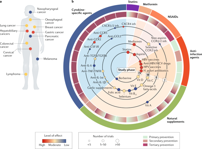
Targeting cancer-promoting inflammation — have anti-inflammatory therapies come of age?

Cancer shared this article with you from Inoreader

41571_2020_459_Fig1_HTML.png

View on the web

Hypoxia-inducible miR-196a modulates glioblastoma cell proliferation and migration through complex regulation of NRAS

Cancer shared this article with you from Inoreader

13402.jpg

Abstract

Background

Glioblastoma (GBM) is the most common and aggressive malignant brain tumor in humans. Hypoxia has been correlated with the aggressive form of glial tumors, poor prognosis, recurrence and resistance to various therapies. MicroRNAs (miRNAs) have emerged as critical mediators of hypoxic responses and have shown great potential for cancer diagnostics and therapeutics. Here, we focus on the regulatory and functional characterization of miR-196a, a hypoxia-inducible miRNA, in GBM.

Methods

Hypoxia/HIF regulation of miR-196a was assessed by RT-qPCR, promoter-luciferase and ChIP assays in GBM cell lines. miR-196a levels were analyzed in The Cancer Genome Atlas (TCGA)-GBM, Chinese Glioma Genome Atlas (CGGA) and Indian GBM patient cohorts. miR-target interactions were studied using RNA/protein quantification and 3'UTR luciferase assays. The effect of miR-196a overexpression/inhibition was assessed on cellular viability, migration and apoptosis under hypoxia and normoxia. Microarray-based gene expression profiling studies were performrd to study the effect of miR-196a on the GBM cellular transcriptome under hypoxia.

Results

We identified miR-196a as a hypoxia-inducible and hypoxia-inducible factor (HIF)-regulated miRNA that plays an oncogenic role in GBM. miR-196a was found to be significantly up-regulated in TCGA-GBM, CGGA glioma as well as Indian GBM patient cohorts. miR-196a overexpression was found to induce cellular proliferation, migration, spheroid formation and colony formation and to inhibit apoptosis, while miR-196a inhibition using anti-miR-196a yielded opposite results, suggesting an oncogenic role of miR-196a in GBM. We further unveiled NRAS, AJAP1, TAOK1 and COL24A1 as direct targets of miR-196a. We also report a complex competitive regulation of oncogenic NRAS by miR-196a, miR-146a and let-7 in GBM. Analysis of microarray-based gene expression data obtained by miR-196a inhibition under hypoxia revealed a role of miR-196a in HIF, calcium adhesion, Wnt and cell adhesion pathways. Interestingly, miR-196a was found to positively regulate the expression of various genes involved in the induction or stabilization of HIFs and in maintenance of hypoxic conditions, thereby suggesting the existence of an indirect miR-196a/HIF positive feedback loop under hypoxia.

Conclusions

Overall, our work identifies a novel association between hypoxia/HIF signalling and miR-196a in GBM and suggests its therapeutic significance.

View on the web

PD-1 and PD-L2 expression predict relapse risk and poor survival in patients with stage III colorectal cancer

Cancer shared this article with you from Inoreader

13402.jpg

Abstract

Background

Immune responses have long been an area of interest in cancer research. In this study, the effects of programmed cell death-1 (PD-1) and its ligand (PD-L2) on the prognosis of colorectal cancer (CRC) were investigated.

Methods

Primary tumour specimens of stage III CRC patients operated between 2002 and 2013 were assessed for PD-1 and PD-L2 expression and various clinicopathological and prognostic factors.

Results

We observed a significant relationship between poor prognostic factors and PD-1/PD-L2 expression. These biomarkers were also found to serve as independent risk factors for LIR and MSI. In univariate analysis, relapse-free survival (RFS) and overall survival (OS) rates were found to be poor in PD-1 and PD-L2 positive patients. In multivariate analysis, these biomarkers were found to serve as independent poor prognostic factors for RFS and OS.

Conclusions

Our data indicate that PD-1 and PD-L2 may serve as independent prognostic survival parameters for CRC patients and may be employed for the design of targeted therapies.

View on the web

Clinical perspectives of BET inhibition in ovarian cancer

Cancer shared this article with you from Inoreader

13402.jpg

Abstract

Background

Bromodomain and extra-terminal (BET) proteins are epigenetic readers that bind to acetylated lysines of histones and regulate gene transcription. BET protein family members mediate the expression of various oncogenic drivers in ovarian cancer, such as the MYC and Neuregulin 1 (NRG1) genes. BRD4, the most thoroughly studied member of the BET family, is amplified in a significant subset of high-grade serous carcinomas (HGSC) of the ovary. It has been reported that BET inhibitors can attenuate the proliferation and dissemination of ovarian cancer cells by inhibiting oncogenic pathways, such as the FOXM1 and JAK/STAT pathways. BET inhibition can re-sensitize resistant ovarian cancer cells to already approved anticancer agents, including cisplatin and PARP inhibitors. This synergism was also confirmed in vivo in animal models. These and other preclinical results provide a promising basis for the application of BET inhibitors in ovarian cancer treat ment. Currently, Phase I/II clinical trials explore the safety and efficacy profiles of BET inhibitors in various solid tumors, including ovarian tumors. Here, we review current knowledge on the molecular effects and preclinical activities of BET inhibitors in ovarian tumors.

Conclusions

BET proteins have emerged as new druggable targets for ovarian cancer. BET inhibitors may enhance antitumor activity when co-administered with conventional treatment regimens. Results from ongoing Phase I/II studies are anticipated to confirm this notion.

View on the web

Protein arginine methyltransferase 5: a potential cancer therapeutic target

Cancer shared this article with you from Inoreader

13402.jpg

Abstract

Background

PRMT5 is a type II protein arginine methyltransferase that methylates histone or non-histone proteins. Arginine methylation by PRMT5 has been implicated in gene transcription, ribosome biogenesis, RNA transport, pre-mRNA splicing and signal transduction. High expression of PRMT5 has been observed in various cancers and PRMT5 overexpression has been reported to improve cancer cell survival, proliferation, migration and metabolism and to inhibit cancer cell apoptosis. In addition, PRMT5 has been found to be required for cancer stem cell survival, self-renewal and differentiation. Several microRNAs have been shown to regulate PRMT5 expression. As PRMT5 has oncogene-like properties, several PRMT5 inhibitors have been used to explore their efficacy as potential drugs for different types of cancer, and three of them are now being tested in clinical trials.

Conclusions

In this review, we summarize current knowledge on the role of PRMT5 in cancer development and progression, including its functions and underlying mechanisms. In addition, we highlight the rapid development of PRMT5 inhibitors and summarize ongoing clinical trials for cancer therapy. By affecting both tumor cells and the tumor microenvironment, PRMT5 inhibitors may serve as effective anti-cancer agents, especially when combined with immune therapies.

View on the web

In silico transcriptomic mapping of integrins and immune activation in Basal-like and HER2+ breast cancer

Cancer shared this article with you from Inoreader

13402.jpg

Abstract

Purpose

Integrins, transmembrane receptors that mediate cell-extracellular matrix and cell-cell interactions, have been linked to several cancer-associated features. A less explored function of integrins in cancer is their role in leukocyte homing and activation. Understanding their relationship with immune cell infiltrates and immune checkpoints is an area of interest in cancer research.

Methods

The expression of 33 different integrins was evaluated in relation with breast cancer patient outcome using transcriptomic data (Affymetrix dataset, exploratory cohort) and the METABRIC study (validation cohort). The TIMER online tool was used to assess the association of the identified integrin genes with immune cell infiltration, and the TCGA and METABRIC studies to assess correlations between integrin gene expression and genomic signatures of immune activation.

Results

We identified 7 genes coding for integrin α and β subunits, i.e., ITGA4, ITGB2, ITGAX, ITGB7, ITGAM, ITGAL and ITGA8, which predict a favorable prognosis in Basal-like and HER2+ breast cancers. Their expression positively correlated with the presence of immune cell infiltrates within the tumor (dendritic cells, CD4+ T-cells, neutrophils, CD8+ T-cells and B-cells), with markers of T-cell activation and antigen presentation, and with gene signatures of immune surveillance (cytotoxic T lymphocyte activation and IFN gamma signature). By contrast, we found that genes coding for integrins that predicted a detrimental outcome (IBSP, ITGB3BP, ITGB6, ITGB1 and ITGAV) were not associated with any of these parameters.

Conclusions

We identified an integrin signature composed of 7 genes with potential to recognize immune infiltrated and activated Basal-like and HER2+ breast cancers with a favorable prognosis.

View on the web